
What Is X-Ray Imaging?
X-Ray Imaging is a diagnostic imaging method that uses a controlled beam of radiation to create pictures of internal structures, especially bones and joints. A detector records how tissues absorb the beam, producing an image that may help a qualified healthcare professional assess alignment, fractures, degenerative changes, or other structural findings.
Quick Facts About X-Ray Imaging
Category
Diagnostic imaging
Used for
Assessing bones, joints, and alignment
Common confusion
An image does not identify every pain source
Also called
Radiography, Plain radiography
Often discussed with
Chiropractic Adjustment, Auto Accident Injury Treatment
Key Takeaways About X-Ray Imaging
- X-ray images show structural detail, especially in bones and joints.
- A qualified professional interprets images alongside your examination.
- X-rays do not directly show every source of pain.
How X-Ray Imaging Works

X-Ray Imaging sends a controlled beam through a body area toward a detector. Dense structures absorb more of the beam and appear lighter, while less dense tissues appear darker. The resulting image shows the shape and position of structures in the selected area.
Related glossary terms: Cervical Spine, Lumbar Spine, Range Of Motion.
Radiography can show bone position, joint spaces, and some changes in spinal structure. A qualified healthcare professional reviews the image with your history and physical examination. The image provides structural information, while symptoms and movement findings add context. X-ray imaging does not directly display every muscle, nerve, or cause of pain.
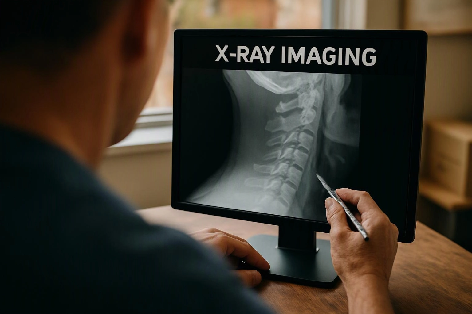
From the Advanced Injury Care Clinic team
When we consider X-Ray Imaging during a chiropractic adjustment visit, we relate the images to the examination and reported symptoms. Our team looks for structural findings that may affect care planning, while recognizing that an image is only one part of the clinical assessment.
X-Ray Imaging in Practice: A Real-World Example
A person with neck pain after a collision may receive X-Ray Imaging as part of an evaluation before a chiropractic adjustment. The images may help assess spinal alignment or visible bone changes alongside the examination.
Related Terms
Cervical Spine
Cervical Spine is the upper section of the spinal column in the neck. It includes the…
Lumbar Spine
Lumbar Spine is the lower section of the spinal column between the thoracic spine and sacrum…
Range Of Motion
Range Of Motion is the available movement at a joint or group of joints, measured through…
Acute Injury
An acute injury is a sudden physical injury that develops after a specific event, such as…
Advanced Injury Care Clinic
Have Questions About X-Ray Imaging?
Contact Advanced Injury Care Clinic for practical guidance on X-Ray Imaging and related chiropractor work in Hendersonville.